

Course Overview:
This Comprehensive Breast Imaging Course, led by László Tabár, MD, FACR, (Hon) and with participation of the Faculty members will offer radiologists the following:
- Normal mammograms will be mixed with proven abnormal cases.
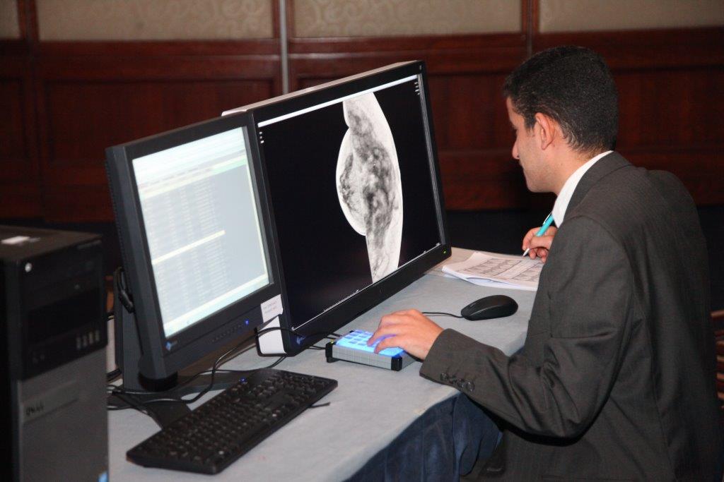
- Reading of normal and abnormal mammograms will take place using an interactive technique (see page III).
- During the course the attendees will progressively improve their interpretive exper-tise, as they learn the full spectrum of normal breast images, with all findings Explained with the help of 3-dimensional histology images.
- These skills will lead to fewer call-backs and greater confidence in reading large number of mammograms.
- Special emphasis will be placed on finding early phase breast cancers.
- All abnormal cases are fully worked up. The complete workup will be presented in detail, including hand-held ultrasound, automated breast ultrasound, MRI and large section histopathology. There will be a special session about tomosynthesis, breast MRI and ABUS (automated breast ultrasound).
- Special sessions will describe the current clinical roles of breast MRI and tomosynthesis, review the image patterns of malignant breast diseases, correlate the findings with the underlying pathology.
- Description of the recent technical advances in breast MRI, including imaging protocols and techniques needed to produce high quality breast MRI images.
- Teaching how to characterize breast lesions utilizing multimodality imaging, breast MRI and tomosynthesis included.
- Learning MRI reading and interpretation at high resolution workstations.
- Studying tomosynthesis cases at high resolution workstations).
Attendees interpreting all interactive digital mammography examinations, hands-on breast MRI, ABUS and tomosynthesis cases will receive a Certificate, confirming that they have read the above mentioned breast imaging cases under the direct supervision of an interpreting physician.
Course Objectives:
- Learn the full spectrum of normal mammograms through detailed explanation of the mammographic images.
- Progressive improvement of the attendees' interpretive expertise.
- Increase confidence in reading large numbers of full field digital mammograms at lower call-back rates.
- Improve skills in detecting early phase breast cancer at digital mammography screening.
- Greater proficiency in working up screen-detected findings.
- Appreciate the clinical relevance of unifocal/multifocal/diffusely infiltrating breast cancers.
- Emphasize the importance of multimodality approach to workup cases in a multidisciplinary environment.
- Assess the clinical role of breast MRI in patient selection and in improving the detection, diagnosis and treatment of breast diseases.
- Characterize breast lesions utilizing multimodality imaging, breast MRI included. The goal is to accurately and efficiently identify, interpret and report on breast MRI examinations


